News
Biosignal Lab Blog
Check out this space for recent news and posts from the Biosignal Lab members.
Ageing and Brain Waves in Big Data
September, 2023
Most neuroimaging studies contain data from tens of participants, which substantially limits the generalizability and reproducibility of the associated findings. Recent trends towards large open-access datasets have provided opportunity to address this weakness. In particular, the CamCAN dataset contains MEG, structural MRI, fMRI and demographic/cognitive data from roughly 600 participants. With an equal distribution of ages from 18-88, this dataset is ideal for cross-sectional investigation of age-related trends.
We have published a series of papers revealing age-related trends in the CamCAN dataset. The dataset includes functional imaging (MEG/fMRI) during a simple cued button press task and during wakeful resting. In particular, we have been looking at the transient signals associated with rhythmic bursts in the sensorimotor cortices. The focus has been on beta band (~20 Hz) transients, although some other signals have been investigated too.
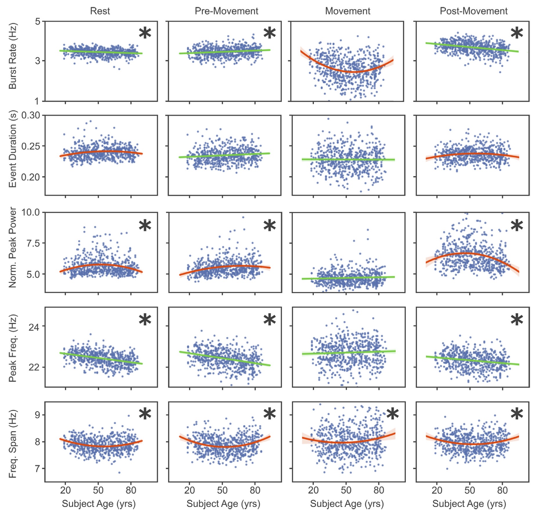
In the cued button press task, we have showed a number of changes in burst characteristics occurring in the contralateral sensorimotor areas. These effects with age include:
- Increasing magnitude of beta suppression
- Likely due to an increase in the pre-movement beta burst rate
- Decreasing peak frequency of beta rebound
- Likely due to a decrease in the peak frequency of beta
- Decreasing amplitude of beta rebound
- Likely due to:
- an increase in the pre-movement beta burst rate, and
- a decrease in the post-movement beta burst rate
- Likely due to:
- Decreasing amplitude of the movement-related gamma burst
- The source of this change has not been investigated
- N-shaped quadratic trend in burst power
- Pre-movement
- Post-movement
- U-shaped quadratic trend in frequency span
- Pre-movement
- Movement
- ±Ę´Ç˛őłŮ-łľ´Ç±ą±đłľ±đ˛ÔłŮĚý
- During wakeful resting we found the following trends,
- N-shaped quadratic trend in burst power
- Linear decrease in burst rate
- Linear decrease in burst frequency
- U-shaped quadratic trend in frequency span
We also developed a new algorithm for generating spatial maps of (and localizing the sources of) transient events (Power, Neuroimage, 2021). This algorithm uses minimum norm estimation to localize a change in spectral power between the pre-burst and burst intervals. MNE was preferred to beamformer for this approach, based on a qualitative comparison of the resulting maps. We showed that beta bursts have different spatial maps in wakeful resting, as compared to pre-movement, as compared to post-movement. In particular, we found an anterior shift in the spatial map for post-movement, as compared to pre-movement, which aligns with previous studies of rebound localization.
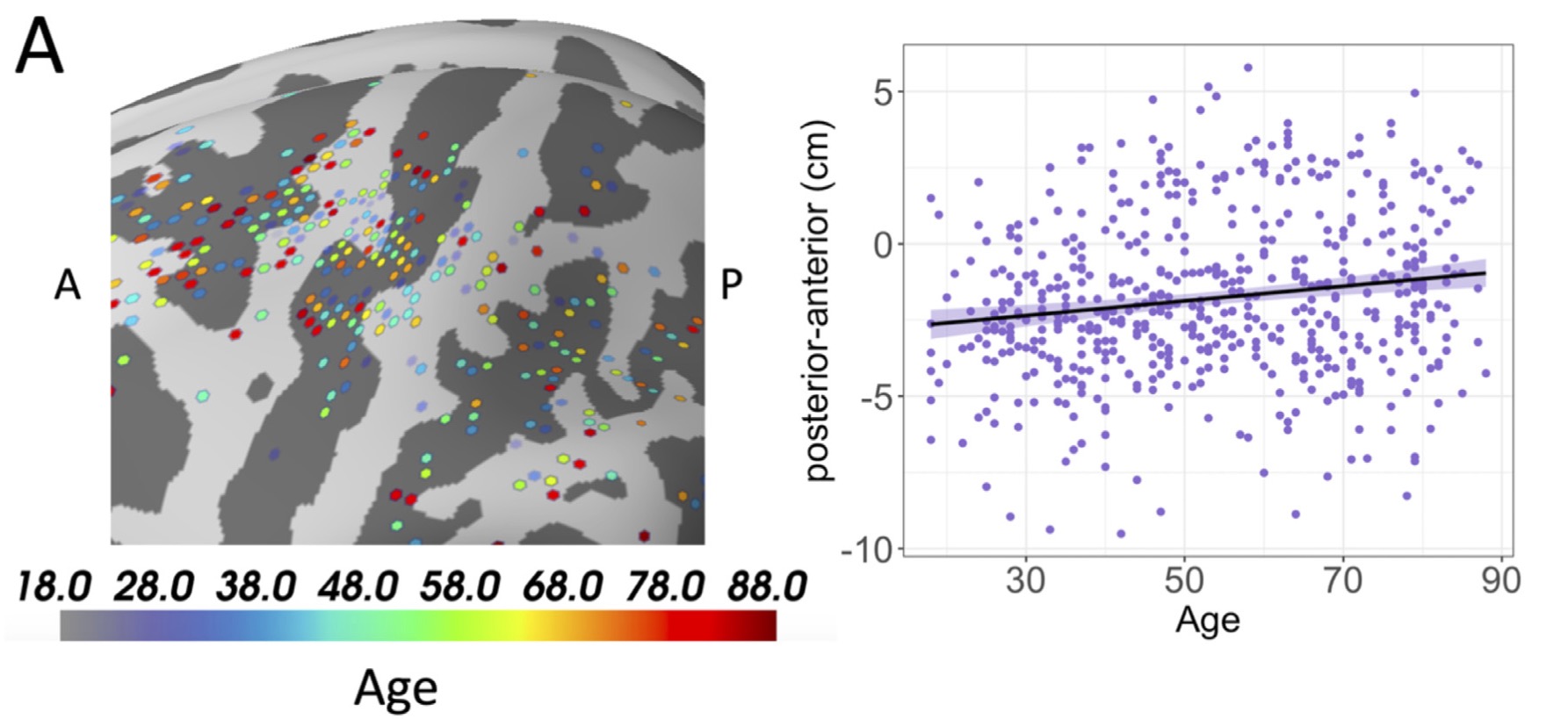
In terms of ageing effects, we found an anterior shift (from contralateral post-central gyrus to pre-central gyrus) in the peak location of the spatial map for the post-movement (i.e., rebound) interval and during waveful resting. As well in the post-movement interval, frontal areas (such as medial and superior frontal gyrus) showed an n-shaped quadratic trend in mean activation with age.
These are just some examples of what can be discovered thanks to the large open-access datasets available online. Here’ I’ve just talked about one type of burst (“beta”) in one area of the brain (primary sensorimotor cortex). There’s so much more to discover here, and we will continue to dig in the big data sandbox!
State of the Slate by Ming Scott
November 9, 2018
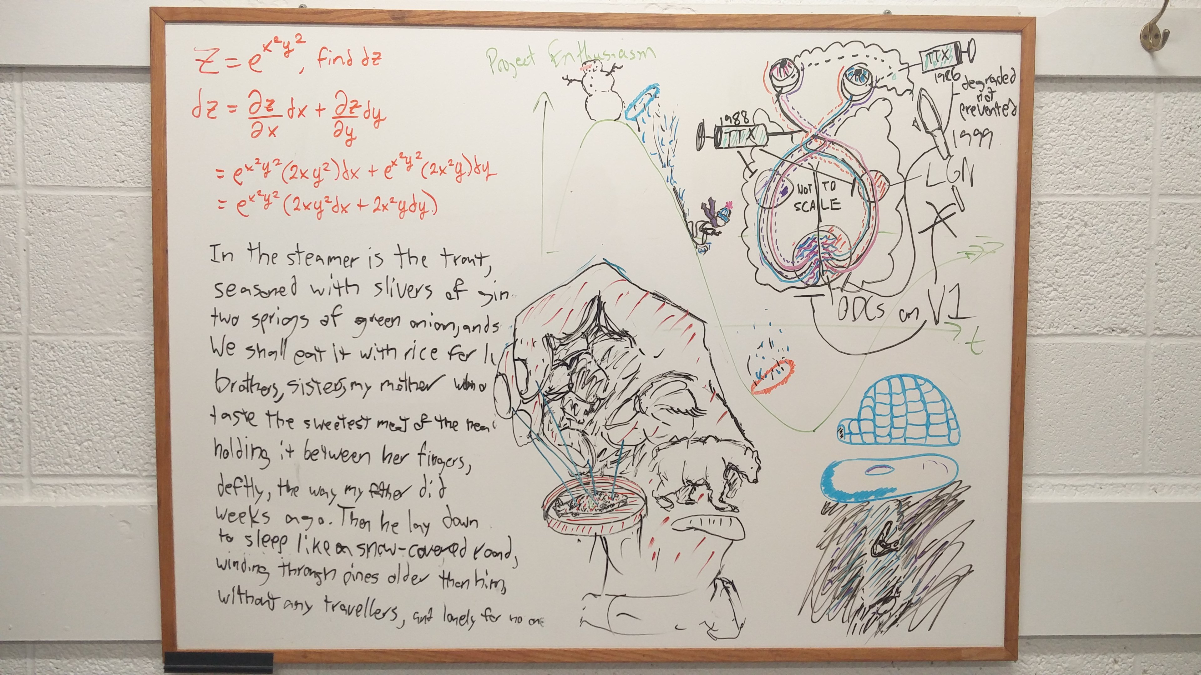
You know it's serious when you stay the night for the first time. Sometimes it's that special someone. Sometimes it's a friend whose couch is always open to you. Sometimes it's a space where you can blast Pink Floyd from a tinny speaker at 3 am, and no one cares that you're tracing out the retinogeniculate pathway in radioactive tints, that you're alternately belting out the approximate lyrics to "Time" and muttering to yourself about tetrodotoxin and retinal waves.
But: I had a paper to finish, I had keys and after-hours access. I had approximately 12 hours until the deadline. I hadĚýpermission.
(Actually, no, I didn't have permission. Hey, Tim, you look at these before they go up. It's, like,ĚýokayĚýto pull drug-fueled all-nighters in the lab, yeah?)
I started at the Biosignal Lab halfway down September, and even though my only duties here are to assist with Lindsey's honours project and water the cacti (yes, really) I find myself spending a lot of time here. It's a good space. It's a friendly space. It's an inspiring space. I come here to study, and the studying comes easy. I come here to write, and the ideas come fast.
The sense you get, walking in here, is that this is a place where people step across boundaries. You don't have to be a particular thing here. It's a physics lab doing neuroscience research, but the space is open to anything you bring to it. It's a place to belong and to be, not just a space to use. People come here to grade psych labs, do stats assignments, write neuroscience papers, memorize poems, to just sit down on the beanbag chair and study.
That's what I like about the whiteboard. The Slate, if I'm staying on-brand. It's a tool for people to think, to relax, to demonstrate, and it's something of a cross-section of the people that use it. I hope people use it as they move through here, wiping away anything in the way. And if they draw something they like, or see something they like, maybe they can take a snapshot and write one of these themselves.
Ěý
Graduate Student Profile - Ron Bishop
Ron Bishop was my first graduate student. He came to ±«Óătv from Paradise, Newfoundland to complete a research project in Rehabilitation Research. His project investigated how brain areas communicate during the performance of precision hand movements. After graduation, Ron worked at the hospital as an Imaging Technician for a number of years. He is now a Data Analyst for the Department of Health and Wellness.
“Getting my masters at Dal was an incredible experience. I learned a lot about neuroimaging and gained so many skills related to research and writing from my supervisors. My time at Dal led directly to an opportunity to work in the field as a technologist, where I collaborated with scientists and met some amazing people."
Where Will Research Take Me? A Review of Recent Grads
For many people, it is hard to imagine where the path of scientific research will lead them. The road straight ahead leads from an undergraduate degree, to a Masters, PhD, post-doctoral fellowship, and finally to a faculty position at a university. Not everyone will make this destination (the tolls are quite high!). But there are many exits from the highway along the way, and those exits can lead to some exciting adventures!
With this in mind, I wanted to share some examples of the paths that some of my previous students have found.
I have supervised a number of honours thesis projects for students in the final year of their undergraduate degrees. For some of these students, this project was their first exposure to research. Others had already developed a passion for scientific discovery. Regardless of their prior experience, they all managed to take on an 8-month project, make it their own, complete the work, and share their findings. I like to think that these experiences helped them build professional skills for success.
At the graduate level, students gain important experience in independent project planning and execution, time management, and in working as part of a professional team. In the hospital environment, they also gain experience interfacing with patients, which can be a very important asset for some career options. Indeed, some of my past graduate students and research associates have left the academic track for promising careers after completing their degree.
Past students – Where Are They Now?
- Data Scientist – Vancouver
- Medical Physics Graduate Student – Halifax
- Data Analyst - Halifax
- Data Scientist – Silicon Valley
- Physiotherapy student – Halifax
- Medical School student – Halifax
Lindsey Presents at North American MEG Meeting
November 8th, 2017
Congratulations to Lindsey Power on her poster presentation at the North American MEG Meeting in Bethesda, Maryland. Lindsey presented some of her work completed during a summer research placement with the Biosignal Lab. Her poster looked at the variability in pre-surgical mapping of brain function between systems made by different vendors.
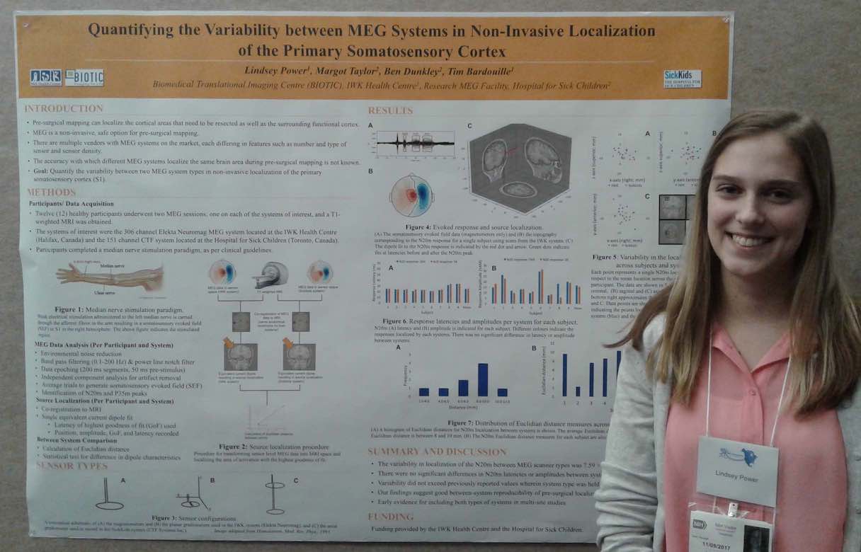
Lindsey's poster was well received and she got lots of positive feedback (plus a trip to DC!).
Great work!
Recording Brain Signals In The Dunn!!!
October 17th, 2017
We had success today recording brain activity with our open-source wireless EEG (electroencephalography) system! This is a big development step towards running studies with wireless EEG in the near future.
Thanks to everyone in the lab who worked so diligently to make this happen. Let's keep moving forward!
Below, you can see power spectra from 8 electrodes placed on the scalp of our healthy volunteer. Data were recorded via bluetooth at 250 Hz while the volunteer rested with their eyes closed.
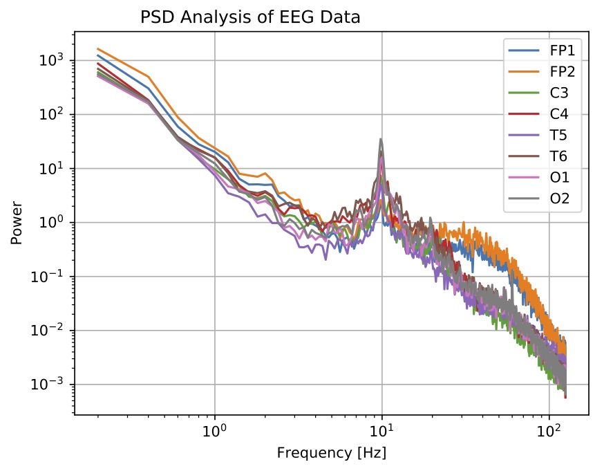
The strong peak at 10 Hz is the "alpha rhythm" first discovered by Hans Berger in the 1930s! You can also see the well-known inverse relationship between so-called background (i.e., non-oscillatory) brain activity and signal frequency. This 1/f relationship is the downward sloping linear tendency seen on this log-log plot. Electrodes FP1 and FP2 have higher signals above 30 Hz, likely due to muscle activity above the eyes because our volunteer was squeezing their eyes shut.
This may be the first time that brain activity has been recorded in the Dunn Building!!!
Physics Meets Neuroscience - Tim Bardouille
October 5th, 2017
People often picture scientists working away in isolation – scribbling notes at a desk or mixing liquids over a flame. In reality, science is very much about being a part of a team. We meet and talk about how we think things work. We work together to figure out how we can test to see if our ideas are right or wrong. Then, we test those ideas experimentally.
It often takes a group of people with different skills to do the experiments. In brain imaging (my line of research), physicists work with physiotherapists, medical doctors, computer scientists, mathematicians, and psychologists, among others. It’s exciting to experience so many perspectives on the same problem.
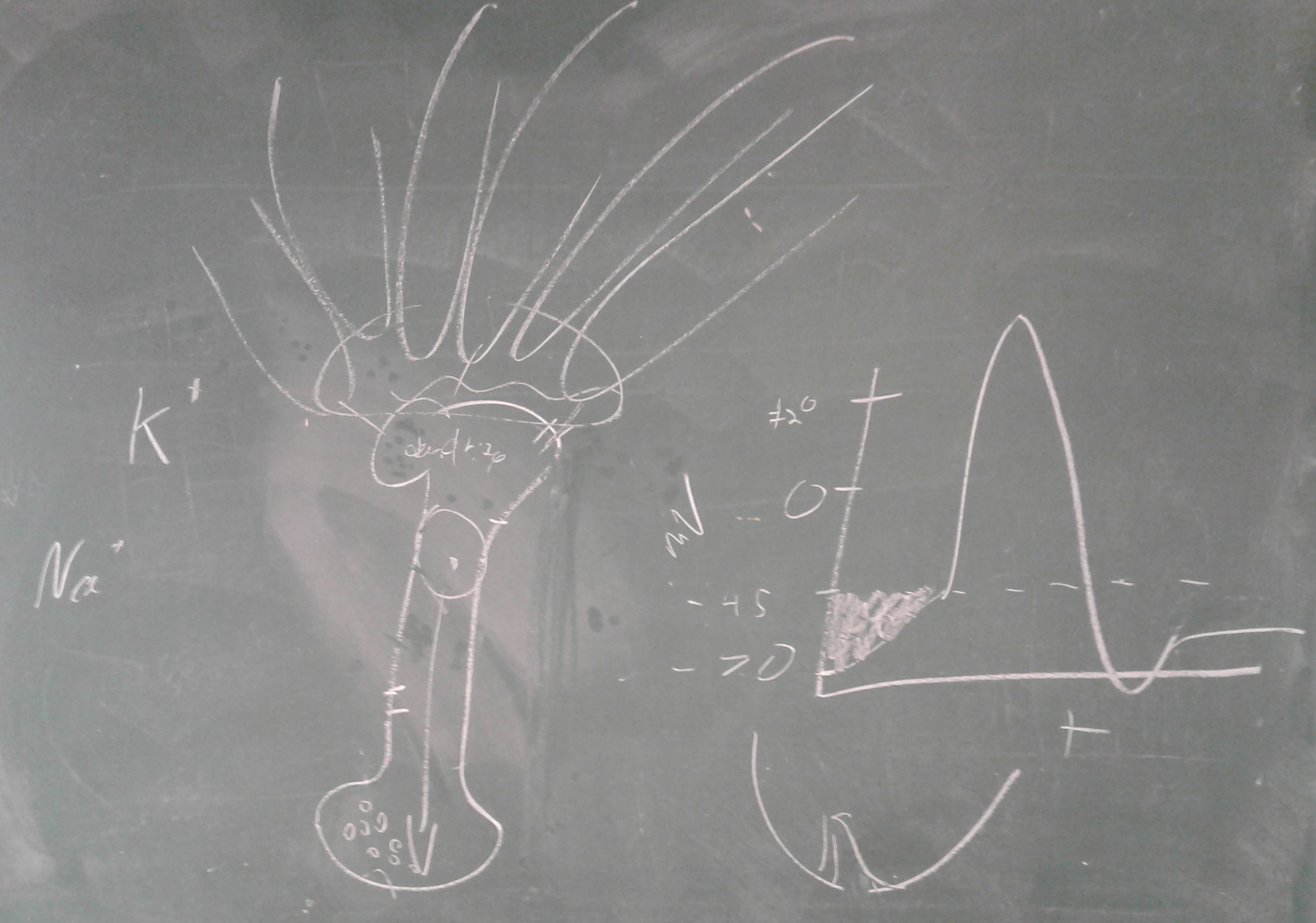
The sharing of knowledge from different areas was really brought home to me when I found this drawing on a chalkboard in my lab. It seems that Justin, a neuroscience undergraduate on my team, was teaching Jon, a Masters student in physics, about how neurons work. ĚýThis is new stuff for Jon, who has worked on other problems in physics in the past. But as a physicist, he already has a deep understanding of what is meant by membrane “potential”!
It’s great to see that scientific discovery isn’t a solitary experience. Ěý
Ěý
Spencer Osbourne Graduates From The Biosignal Lab
August 30, 2017
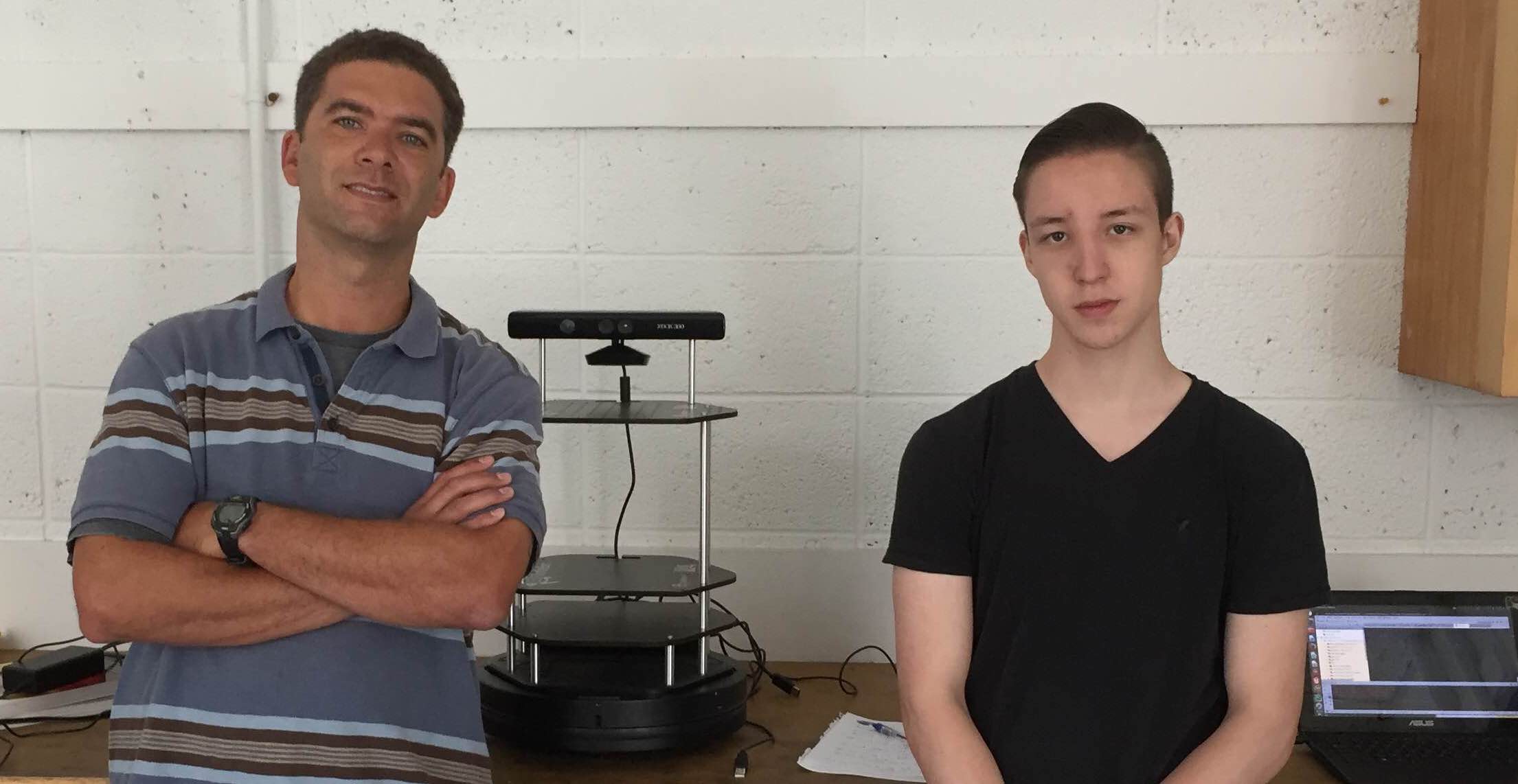
Thanks to Spencer Osbourne, who spent the summer with us at the Biosignal Lab. Spencer is beginning his last year of high school, and has volunteered with me the last two summers.
This summer Spencer developed some code to help our robot navigate around a room without bumping into things. He did a great job on this project, which will now be taken up by some interested students in the Physics department.
Great to have you on the team! Thanks, and good luck with Grade 12!!!
